


About Us
Executive Editor:Publishing house "Academy of Natural History"
Editorial Board:
Asgarov S. (Azerbaijan), Alakbarov M. (Azerbaijan), Aliev Z. (Azerbaijan), Babayev N. (Uzbekistan), Chiladze G. (Georgia), Datskovsky I. (Israel), Garbuz I. (Moldova), Gleizer S. (Germany), Ershina A. (Kazakhstan), Kobzev D. (Switzerland), Kohl O. (Germany), Ktshanyan M. (Armenia), Lande D. (Ukraine), Ledvanov M. (Russia), Makats V. (Ukraine), Miletic L. (Serbia), Moskovkin V. (Ukraine), Murzagaliyeva A. (Kazakhstan), Novikov A. (Ukraine), Rahimov R. (Uzbekistan), Romanchuk A. (Ukraine), Shamshiev B. (Kyrgyzstan), Usheva M. (Bulgaria), Vasileva M. (Bulgar).


Materials of the conference "EDUCATION AND SCIENCE WITHOUT BORDERS"
 PDF
PDFThe coral – type and multiple nephrolithiasis (СMN) is the severe form of the kidney stone disease (KSD). The emergence of the new medical treatment and therapies methods KSD have already been entered and have been made the specific certain changes in this process, and more and more supporters of the minimally and low invasive medical treatment methods (1,2).
However, when the large coral – like calculi’ (CC) are being filled the pelvis, all groups of the cups, and, at the same time, having had the narrow neck messages of the messages are reached in the cups, relatively, the large sizes, of course, the open surgery has its advantages.
Thus, in the period of 1998 to 2012, we have already operated the 2,050 patients with the kidney calculi. The 730 (e.g. 35,6%) ones have been with СMN (480 (65,7%) patients have been with the one – sided, 250 (34,2%) patients with the doubled – sided coral – type calculi’) of these ones.
The patients’ survey examination has been conducted on sick people with the common and generally accepted tactics, and it has been included the general blood and urine analyses, the ultrasound and X – ray examination, and to be determined the CRL stage they have performed the Tehberg – Tareev test. In those case, when there the contra – EU, the spectral CT is the choice method for the diagnostics. Before the surgery operation conduction, all patients have been underwent the intensive conservative medical treatment.
The CMN patients have been ranged in the age from 4 up to 76 (e.g. the average age 49 years). The total males have been 351 (e.g. 48.0%), the females – 379 (e.g. 51.9%). From the observed and the examined ones, the CC have been combined with the multiple calculi’ at 440 (e.g. 60,2%), and the only CC have been at the rest – 290 (e.g. 39,8%) patients.
The stage of the disease has been determined at the observed and examined patients by the CN classification in shape and size, having adopted at the USSR All – Union Congress of Urology in 1990 (6).
The work’s objective: to be studied the results for the open surgery operations at the patients with the СMN, having conducted with the renal arteries‘ cross – clamping, against the background of the anti – ischemic protection of the kidney.
To be removed the calculi’, we widely used, as the pyelotomy, well as nephrotomic cuts. Here, one of the basic highlights is to be ensured the minimal loss of the blood provision. Therefore, during the operation conduction, the temporal renal ischemia, due to its vascular cross – clamping and also its anti – ischemic protection, has the significant and significant value. So, the renal blood flow cross – clamping and its overlapping during the period of the intervention is removed the threat of bleeding, although it is associated with the renal ischemia. The organ’s ischemic injury is usually presented itself the cascade process, having deeply affected all the entire cell’s structure. The existing ways of dealing with the ischemic stress are allowed only partially to be protected the kidney from its following changes, having occurred in it during the period of the ischemia and its reperfusion. However, the basic steps to be prevented the organ’s ischemic injury consequences is the affordable realty. So, in the practical operative urology, to be solved this challenge has been found for their application: the local renal hypothermia, as well as its pharmacological protection.
So, it is no the secret, that at the local hyperthermia method using, one of the main conditions for the following success is the preliminary administration of the mannitol (e.g. 12,5–25g.), which is reduced the intra – canalicular crystallization during the hypothermia, and it is prevented, moreover, the intracellular edema. Thus, this method is, besides cumbersome, than the technical complexity, is much more needed yet in the additional medical treatment of this preparation (e.g. mannitol), having possessed by its anti – ischemic medical properties.
In its turn, the pharmacological method, because of its simplicity, affordability and efficiency, that does not require the technically complex manipulations, is practically more attractive, in order to be protected the kidney from the ischemia.
For more already more, than 20 years, at the operative surgical interventions on the kidney, having passed with the cross – clapping and overlapping the renal blood flow, so in this aspect, such as, we have already successfully applied the furosemide (e.g. Lazix) (3). Having acting from the canaliculus lumen’s side, it selectively is inhibited the sodium combined transport system in the canalicular epithelium cells. This is lead to the energy consumption number reduction and the ATR accumulation in the cells, resulting in the kidney tissue resistance is increased to the hypoxia. Then, the furosemide is also increased the renal blood flow, it is caused the blood redistribution in the kidney cortical layer, and it, moreover, is contributed the hypo-coagulation.
At the same time, having taken into account the significant role of «the calcium paradox» and the «no-reflow» phenomenon in the expressed post – ischemic disorders development, we have already used the calcium canals’ blocker – the verapamil (e.g. Izoptin), in the conjugation with the furosemide more, than 10 years (e.g. 120 months). The calcium canals blockers, in particular, the verapamil, having stabilized the membranes, are practically protected the cell from the calcium accumulation, and they, moreover, are prevented the post – ischemic arteriolar spasm development. The conducted in the recent years, numerous studies have been shown, that in the organ’s ischemic injury, along with the cell’s ionic balance violation, the major role is played by the lipid peroxidation (LPO) activization. It has been convincingly proved and shown, the sharp increase in the LPO products content in the reperfusion period in the ischemic organ’s blood. The LPO activization is practically resulted in the severe injury membranes and in the cell’s energy with its vital activity of the life violation. Having guided by the above mentioned, we have experimentally investigated and introduced into the clinical practice the antioxidant action preparation for the complex introduction with the furosemide and the verapamil in the struggle against the kidney’s ischemic and the operational stress. In this aspect, this preparation is quite useful, as the drug, which could be administrated directly into the bloodstream, as on the operating table, well as in the next early post – operative period. Undoubtedly, along with the antioxidant effect, it should be also possessed and other properties and characteristics, which are quite useful against the struggle with the kidney’s ischemic injury. This preparation and the corresponding drug has been become emoksipin (4). To be protected the kidney from the ischemic injury, at the intra-operative cross-clamping renal artery, the furosemide and the verapamil in the doses 3 mg/kg and 0,2 mg/kg, and also the emoksipin (e.g. 1 mg/kg), respectively, are administrated 15 minutes prior to the a.renalis cross- clamping and, immediately, after the renal blood flow restoration. Also, all these preparations and the corresponding drugs, at the same doses, are being administrated to the patient within the 5 post-operative days (e.g. 120 hours), to be created the improved kidney’s function. In the recent years, for the kidneys’ anti – ischemic protection, we have already, experimentally, investigated the efficiency, and have also begun to be used the perftoran clinical application (5).
Here we are watching the successful surgery intervention at the patient with СMN.
The patient S.S., a male of 57 years old, medical patient’s history № 192, has been admitted to the hospital 05.12.2012, with the pains in the lumbar region and the overall asthenia. The clinical diagnosis: «The coral–type and multiple calculi’ of the left kidney. The hydronephrosis is on the left. There is the chronic renal insufficiency (in the stage of compensation)».
The Laboratory Studies. The Complete blood count (CBC): Hb 106q/l, eritr. 3,6 x 10¹², leyk. 9,5 x 10⁹, ESR 25 mm/h.
The general analysis of urine: the acidic reaction, the relative density – 1,006,
the protein – 0,5q/l, the white blood cells are completely covered the field of view. The Zimnitzky test is given the urine’s relative density – 1,010 – 1,015.
USS. The dimensions of the right kidney – 11,8 x 5,9sm, the parenchymal thickness – 1,5 sm. The dimensions of the left kidney – 12,8 x 6,1 sm, the parenchymal thickness – 1,4sm, the coral – type calculus is revealed, measuring – 3,5 х 3,2 х 1,2 х 1sm.
At the Review Voiding in the projection of the left kidney, at the level of the LI-III vertebrae, the coral-type calculus shadow is defined (fig. 1a). On the excretory urogram, in the 60-th minute, the both kidneys’ function is satisfactory, the coral-type calculus is filled the CHLS of the left kidney.
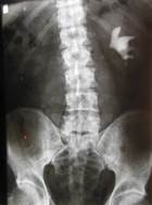

Fig. 1. The Patient S.S.
a. the Review Voiding; b) the Excretory urogram in the 60-th minute.
On 06.12.2012 the patient, under the endotracheal anesthesia, «The Back Sub-cortical Pyelolithotomy Is Left. The Internally Kidney’s Stenting» has been performed. During the operation, it has been established. That there are the expressed cicatricial changes: the kidney is throughout adherent to the cicatricial – changed perirenal fat, resulting in the pelvis has already been taken the intra-renal position. So, the kidney has been very tense. Basically, by sharp, the kidney has been mobilized; the renal artery has been allocated. The back wall of the pelvis has been above the UPJ over 3 cm long and 2,5 cm wide. Obliquely – cross cut 2 cm above the pelvis segment has been opened.
Here, it was found, that the calculus end had been hammered into the segment. To be removed the calculus, so as not to be damaged the segment, firstly, the renal artery has been cross – clamped. The kidney’s tension has been significantly decreased. The calculus end has been hammered into the UPJ. To be avoided the segment’s damage, the CC has been pinned, by its possible inside the glasses, which it has been allowed to be brought the calculus end to the wound. After that, the main and large CC has been removed (Fig.2). The removed calculi’: the CC has 4 branches, dark in color, sizes 6,0 х 4,0 cm. Next, from the kidney, two small calculi’, which size of 1,0 х 0,8 cm., have been also removed.
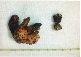
Fig. 2. The Removed Calculi’.
The renal artery’s cross – clamping time has been made up 11 minutes. The pelvis incision has been sutured by the catgut 3/0 on atraumatic needle. The bleeding has not been observed, either during, or after the surgery operation. During 5 post-operative days (e.g. 120 hours), we have conducted the anti – ischemic kidney’s protection, according to the above – described method. The patient has been discharged from the hospital under good health condition on the 12-th days (e.g. 288 hours). The patient has been done the EU after the stent removal (Fig.3). So, it has been done after 8 months (e.g. about 240 days) after the surgery operation. The calculi’ have not been on the USS and on the overview image or the survey picture. The kidney’ excretory function has been adequate, on the left, in comparison with the right side, the CHLS slight expansion has been shown.
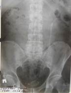
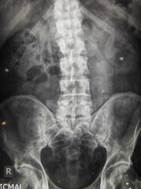
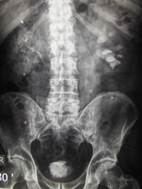
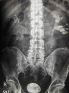
Figure 3. The S.S. patient’s excretory urography or pyelography throughout the 8 months (e.g. about 240 days) after the surgery operation.
a.b. The voiding survey; c.d. The Excretory urogram at the 30-th and 60-th minutes.
The 1,065 surgery operations have been conducted at the 740 СMN patients (e.g. the right side 564, the left side 501). So, the operations have been resulted in the right nephrostomy overlapping at 116 ones (e.g. 15,8%), on the left at 93 ones (e.g. 12,7%), the right pyelostomy at 46 ones (e.g. 6,3%), on the left at 31 (e.g. 4,2%) patients, in the right intrarenal stenting at 181 (e.g. 24%), on the left one at 167 (e.g. 22,8%) patients, the intrarenal stenting and the right nephrostomy at 29 (3,9%) ones, on the left at 6 (e.g. 0,8%) patients. So, the nephropexy has been performed at 210 (e.g. 28,7%) patients at the end of the surgery operation. Of these, at 78 ones (10,6%) have been accomplished by, in the connection of about the accompanying nephroptosis, at 132 (e.g. 18,0%) patients, in the connection of about the forced mobilization(s) of the kidney during the surgery operation.
The nephrectomy has been conducted at 40 (e.g. 5,4%) patients. The cross-clamping renal surgery operation has been performed at 184 (e.g. 25,2%) patients. The right renal artery has been occluded at 80 (e.g. 10,9%) ones, on the left – at 104 (14,2%) patients. So, the operation nature character has been the following: the sub-cortical back pyelolithotomy has been made on the right at 35 (e.g. 4,7%) ones, on the left – at 60 (8,2%) patients. The nephrolithotomy has been performed on the right at 45 (6,1%) ones, on the left – at 60 (8,2%) patients. Thus, the cross – clamping time of the renal artery has been made up, in average, 16,5 (e.g. 5 – 50) minutes.
The average duration of the surgery operation has been made up 2,6 (e.g. 1,5 – 6) hours, the blood loss – 110 (50 – 300) ml, the 500 ml of the blood has been handled at 14 (e.g. 1,9%) patients before the operation, in connection of the anemia (e.g. нв – 75g/l). There has been the chronic pyelonephritis exacerbation has been observed in 40 (e.g. 5,4%) cases, in 25 (e.g. 3,4%) ones – the chronic renal insufficiency, in 25 (e.g. 3,4%) – the wound abscess.
The residual calculi’ have been revealed at 175 (e.g. 23,9%), with the repeated X – ray and the ultrasound examinations before the discharge from the hospital. The observations have been revealed the residual calculus for 1 – 10 years at 238 (e.g.32,6%) operated patients. In these cases, substantially, ESWL has been used for the medical treatment of smaller calculi’. Of them, at 100 (e.g. 13,6%) patients, the surgery reoperation has been underwent.
The Conclusion: In the recent years, the preference tendency of what, and type – method of the medical treatment at СMN without any objective reasons is observed. The principles of the СMN modern medical treatment are required the low – impact, body – survival conducting, the operations with minimal blood loss from the urologists. The quite different and various complications in the form chronic pyelonephritis, the total hematuria, the residual calculi’ formation can be experienced and also observed after the СMN surgical medical treatment. Despite of all these complications, the opened surgical treatment with the anti – ischemic protection observance at the СMN is created the conditions for the safe and thorough kidney’s inspection for the calculi’ removal.
The surgical operating equipment facilities on the removed calculi’ control can be reduced the residual calculi’ number to its minimum. Thus, the proper clinical evaluation, the preoperative preparation, the rational choice of the surgical tactics, the highest vocational and occupational level of the urologist is practically allowed to be optimistically considered the role of the open operations at the СMN.
2. Olefir Yu.V., «The Low-Invasive Treatment Methods of Nephrolithiasis Complex Forms: The Absract of Thesis. The Doctor of Medical Sciences». – Moscow, 2008. – p.p. 52;
3. Imamverdiev S.B. ,«The Surgical Treatment of Coral – Like and Multiple Nephrolithiasis». Baku, 1993., p.p. 107;
4. Imamverdiev S.B., Mamedov R.N., “Emoksipin in Complex Pharmocological Kidney Protection from Ischemic and Prerational Stress(es)”. // «Urology». М. «Medicine», 2003, № 5, p.p. 40 – 42;
5. Imamverdiev S.B., Godjaev M.A., Talybov M.A., Gusein – Zadeh R.T., Mamedov R.N., Nagiev R.N. ,“Perftoran, as the Anti – Ischemic Kidney Protection Choice Method”. «The Experimental Investigation. The Healthy Men». 2011, № 4 (39). р.p. 148 – 150;
6. Yanenko E.K., Khursev K.B., et.al.,,«The Treatment of Coral – Like Nephrolithiasis, Depending on the Disease Stage: The Methodological Recommendations». – М., 1995.
Imamverdiev S.B., Talybov T.A., Mamedov R.N. THE ANTI – ISCHEMIC PROTECTION FOR OPEN OPERATIONS, OVER
THE CORAL-LIKE AND MULTIPLE NEPHROLITHIASIS
. International Journal Of Applied And Fundamental Research. – 2013. – № 2 –
URL: www.science-sd.com/455-24387 (20.04.2024).