


About Us
Executive Editor:Publishing house "Academy of Natural History"
Editorial Board:
Asgarov S. (Azerbaijan), Alakbarov M. (Azerbaijan), Aliev Z. (Azerbaijan), Babayev N. (Uzbekistan), Chiladze G. (Georgia), Datskovsky I. (Israel), Garbuz I. (Moldova), Gleizer S. (Germany), Ershina A. (Kazakhstan), Kobzev D. (Switzerland), Kohl O. (Germany), Ktshanyan M. (Armenia), Lande D. (Ukraine), Ledvanov M. (Russia), Makats V. (Ukraine), Miletic L. (Serbia), Moskovkin V. (Ukraine), Murzagaliyeva A. (Kazakhstan), Novikov A. (Ukraine), Rahimov R. (Uzbekistan), Romanchuk A. (Ukraine), Shamshiev B. (Kyrgyzstan), Usheva M. (Bulgaria), Vasileva M. (Bulgar).


Medical sciences
 PDF
PDF«Movement activity» usually is considered not only as muscular loads - hyperdynamy and hyperkinesia, but also as their restriction - hypokinesia. Physical strain, acting on the locomotor system, has the ability to affect all body functions [1, 2]. Movement activity is accompanied by morphological rearrangements in the cardiovascular, respiratory, endocrine, digestive and nervous systems. However, limited physical activity is now an integral part of human existence [5, 6]. The nervous system is the central part of adaptive changes implementation in the organism under the action of various movement activities. Nowadays the questions relating to the central nervous system are well enough described in the literature, but information about changes in the morphogenesis of the peripheral nervous system structures, namely about features of conducting and stromal components of the peripheral nerves, is not represented in its entirety [7]. The current state of the question has served as an occasion for studying the given problem.
The aim of the study: to investigate the morphological features of the stromal component of peripheral nerves from the upper limbs of experimental animals - rats - under hypo- and hyperkinesia.
Materials and methods
The experimental work was carried out on 72 male rats Wistar, weighing 120-140 at the beginning of the experiment, and 230-250 at the end of the experiment. Treatment of animals, their maintenance and killing was performed in accordance with the European Convention for the Protection of Vertebrate Animals used for Experimental and other Scientific Purposes, adopted by the Council of Europe (Strasbourg, 18 March 1986), and according to the rules of Good Laboratory Practice of the Russian Federation (Ministry of Health order № 267 from 10.06.2003). In order to standardize experimental conditions we chose laboratory male rats as the object of observation. These animals appropriately react to the increase and decrease of movement activity. Animals were kept in a vivarium, where they passed the quarantine and vaccination. All experimental rats have been divided into 3 groups. Animals in the first group were kept under hypokinesia and were subdivided into 9 subgroups, depending on the timing of removal from the experiment. The hypokinesia conditions were created by placing the animals in hypokinetic chambers (patent №82085 from 20.04.2009) [3, 4]. Animals of the second group has also been subdivided into 9 subgroups and contained in conditions of hyperkinesis, which were created by a daily placing of laboratory animals for 3 hours in warm (t 38-40°С) water. The third group was the control. Animals were removed from the experiment by euthanasia under ether anesthesia by decapitation. All experiments with motor load and taking the experimental material were carried out in the afternoon (from 15.00 till 18.00).
The object of this study was the forelimbs of laboratory rats. Upper extremities were dissected at the shoulder joint, after that humeral bones with surrounding muscles were separated to the elbow joint. Further the bone was subperiosteally separated from the adjacent muscles by using universal microraspater (certificate for a reasonable proposal № 1762-07 from 20.05.07). The received material was fixed in 10 % solution of neutral Formalinum. Then transverse paraffin sections with the thickness 10-12mkm were prepared and stained with hematoxylin-eosin by a reference technique.
Results and discussion
As a result of the conducted research the following data were obtained: at preparation the peripheral nerves of the forelimbs two nerves - flexors and one nerve - extensor were allocated on the inner surface of the brachium (Fig.1).
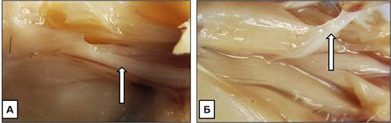
Fig.1. Macroscopic picture of the peripheral nerves of the brachial plexus in the middle third of the shoulder on the medial (A) and lateral (B) surfaces.
The morphologic study found, that on a transversal cut in the top third of brachium the peripheral nerve consisted from 5 on the right and 6 at the left well distinguishable nervous trunks; in the middle third - the "nerve-extensor" both at the left and on the right comprised of three fascicles of a different diameter covered with common fascial vagina. "Nerves - flexors" consisted of 3-4 fascicles of a different diameter, without common fascial vagina, but covered by well-expressed perineurium. In the lower third of brachium the "nerve - flexor" comprised of two large and one small fascicles, and "nerve-extensor" consisted of four small fascicles (Fig.2).
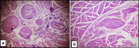
Fig.2. Micrograph of neurovascular bundle with "nerves - flexors" (A) and "nerves-extensors" (B) in the middle third of the shoulder. Increase x 40. Stained with hematoxylin-eosin.
All nervous fascicles were covered by perineural cuff, well identified in layers, consisted of a friable fibrous connective tissue. Moreover, in perineurium it was possible to separate two layers. The first, external, comprised of connective tissue with loosely located fibers and small nervous trunks with blood vessels, disposed in these fibers. The second, internal - perineurium, that had brightly oxyphilous color. It was formed by ordered, closely spaced fibers and covered on the side of nervous fibers by epithelioid, flat cells (Fig.3A). Nervous fascicles were formed by myelin and non-myelin nervous fibers, between them loose fibrous connective tissue interlayer of endoneurium was located. In its structure except fibroblasts there were mast cells at different stages of functional activity (Fig.3B).
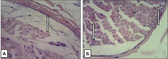
Fig.3. Micrograph of a nerve bundle, surrounded by perineurium (A) and formed by myelin and non-myelin fibers, between which the interlayer of endoneurium is located (B). Increase x 100. Stained with hematoxylin-eosin.
Outside fascicles of nervous fibers were covered by epineurium, contained all cellular elements of a friable fibrous connective tissue, nervous fibres, nervous terminals and blood vessels. In a middle third of brachium the nerve-flexor and being with it in epineurium main vessels on both sides were surrounded by muscles and on the third side - by skin. The nerve-extensor in a middle third of brachium on one side was adjacent to the bone by a layer of loose connective tissue, and on the other side was covered with muscles.
In the experiment, we assessed the stromal component of peripheral nerves. In the period of hyperkinesia on the 7th day there was a thickening of perineurium and its moderate separation from nervous fibres. The thickness of endoneurium was increased in nervous trunks of small diameter, and there were predominantly mast cells of II type (light cells, with well distinct nucleus, loosely filled with pellets) and of III type (light, empty cells with well distinct nucleus and single pellets in cytoplasm) in it. In perineurium mast cells of I type prevailed (dark cells with separately distinct pellets, nucleus is not visible) and also there were brown adipose tissue cells. Mast cells of 0 type (very dark cells, densely filled with pellets, the nucleus is indiscernible) met less often. The reaction of a connective tissue was more expressed by the 14th day. In epineurium in the field of view there were a large amount of lymphocytes, eosinocytes, band and segmented neutrophils, fibroblasts and mast cells mainly of II and III types. In endoneurium the number of fibroblasts and fibrocytes was increased. It is interesting to note, that the cellular structure was more various and brightly expressed in epineurium and perineurium of the nerve-extensor. By the 21th day of experiment reactive stromal component changes were insignificant, but the brightly expressed hyperchromia of lymphocytes nuclei paid attention to itself. By the 30th day of hyperkinesia the perineurium of nerve-extensor was significantly thickened; the general fascial vagina of the nerve was well expressed, in comparison with the previous period of experiment. In the blood vessels of endoneurium the stasis was observed. In nervous fibers the prevalence of thickness myelin shells above the diameter of the axial cylinder was obviously expressed.
On initial terms of hypokinesia the connective tissue fibers were located more compactly, in comparison with the same terms during the hyperkinesia. In epineurium there were plenty of white and brown adipose tissue, cellular structure had less diverse composition. However by the 14th day of hypokinesia similar reactive changes of cellular structure were observed, as it was with hyperkinesia. Mast cells were mainly of 0 and I types. In the blood vessels the stasis was observed. On later terms fibers of epineurium and perineurium became more compact. They were brightly expressed and structured. The amount of mast cells of I and II types, plasmocytes and lymphocytes considerably increases by the 21th day of experiment. It is interesting to note, that axial cylinders of nervous fibers in fascicles became thicker and their diameter prevails on width of myelin shell.
Conclusion
Thus, hypo- and hyperkinesia conditions cause to the structural changes in the stromal component of peripheral nerves of the brachial plexus. The severity of these changes had a direct proportionality to the duration of the experiment. The most pronounced changes were observed in the cellular component of the nerve trunks stroma, the degree of their reactivity is better manifested under hypokinesia. The revealed changes in the connective tissue component of peripheral nerves upon exposure on the organism various regimes and duration of movement activity have expanded representations about adaptation mechanisms of peripheral nervous system to the impacts of various environmental factors. Our findings can be used in neurology, neurosurgery, traumatology and sports medicine.
2. Kataev A.V., Gizatullin T.R. The impact of benzimidizol derivative with dioksotietan cycle on the consequences of exercise-induced emotional stress in animals. // Kazan Medical Journal. - 2015. - Vol.96. - №1. - P. 56-60.
3. Kurbonov Z.A. Treatment of damage aftermath of the neurovascular bundles from upper extremities. Dissertation synopsis for the degree of Medical Sciences Candidate. - Dushanbe, 2006. - 23p.
4. Morozov V.I. et al. Morphological and biochemical aspects of injury and regeneration of skeletal muscles during physical exertion and physical inactivity. // Morphology. - 2006. - Vol. 129. - № 3. - P. 88-96.
5. Vasjagina T. I., Petrova N.I. Change of nerve fibers in the sinuauricular area of the heart and in the thyroid gland after a single maximal exercise and hypokinesia. // Morphology. - 2009. - Vol. 136. - № 4. - P. 29.
6. Zatolokina M.A. et al. The morphofunctional state of connective tissue component of peripheral nerves from experimental animals’ forelimbs after changing their motor activity. Materials of the international scientific conference dedicated to the 65th anniversary of the Ivanovo lymphologists’ school "Actual problems of theoretical, experimental and clinical morphology." Ivanovo, 2009. - P. 154-157.
7. Zatolokina M.A. et al. Structural and functional features of connective tissue component of certain organs after physical activity changing. // Morphology. - 2009. - Vol. 136. - № 4. - P. 61.
Zatolokina M.A., Gerasimova A.V., Tsaroeva L.K., Maleeva D.S. THE MORPHOFUNCTIONAL STATE OF CONNECTIVE TISSUE COMPONENT OF PERIPHERAL NERVES FROM EXPERIMENTAL ANIMALS’ FORELIMBS AFTER CHANGING THEIR MOTOR ACTIVITY. International Journal Of Applied And Fundamental Research. – 2015. – № 1 –
URL: www.science-sd.com/460-24760 (20.04.2024).