


About Us
Executive Editor:Publishing house "Academy of Natural History"
Editorial Board:
Asgarov S. (Azerbaijan), Alakbarov M. (Azerbaijan), Aliev Z. (Azerbaijan), Babayev N. (Uzbekistan), Chiladze G. (Georgia), Datskovsky I. (Israel), Garbuz I. (Moldova), Gleizer S. (Germany), Ershina A. (Kazakhstan), Kobzev D. (Switzerland), Kohl O. (Germany), Ktshanyan M. (Armenia), Lande D. (Ukraine), Ledvanov M. (Russia), Makats V. (Ukraine), Miletic L. (Serbia), Moskovkin V. (Ukraine), Murzagaliyeva A. (Kazakhstan), Novikov A. (Ukraine), Rahimov R. (Uzbekistan), Romanchuk A. (Ukraine), Shamshiev B. (Kyrgyzstan), Usheva M. (Bulgaria), Vasileva M. (Bulgar).


Biological sciences
 PDF
PDFIntroduction.There are significant morphological and functional changes in the whole body, and especially in uterus during the pregnancy and childbirth [1,6]. High-quality prenatal care and the childbirth effective management are directly dependent from morphological and functional changes in cervix and uterus lower segment. The cervix functional morphology investigating experimental studies on mammals can help in practice, to prevent preterm cervical ripening in pregnancy or to be the cause of immaturity at the onset of labor.
The purposes of research was study the collagen type III localization in cervix of intact rats and clarify the changes of its expression in maturing and mature cervix during pregnancy and labor. There were following tasks to achieve this goal:
1. To determine the localization of type III collagen in the cervix nulliparous mature rats;
2. To compare the features of type III collagen expression in intact, maturing and mature cervix.
Material and methods. We used 30 laboratory rats weighing 200-300 g. in our work. The animals were divided on 3 groups: the first (control) group included mature nulliparous no pregnant rats (n = 6), the second group consisted of pregnant females at 15,17,19,20, 21 day of pregnancy (n = 20), the third group consisted of 4 rats during physiological labor. Our experimental studies were carried out in accordance with the "Guidelines for the Care and Use of experimental animals", as well as compliance with the rules of humane treatment of animals (Report of AVMA Panel on Eutanasia IAVMA, 2001 [4]).
We used optical microscopic and immunohystochemistry using antibodies to collagen type III methods in our work. The material was fixed in buffered formalin and then it was carried out in a closed histological processor type vacuum Leica ASP 300. After the material was processed with «Histomix» paraffin (Bio Optica). Frontal, sagittal and transverse sections were prepared on a rotary microtome of 4 microns thickness. The prepared sections were stained with hematoxylin and eosin and Masson method. We used DACO antibodies in our immunohystochemical studies. The immunohystochemical reactions were performed with one-step visualization system BioGenex (QD 630 - HAQ) Super Sensitive one-step Polymer- HRP Kit / DAB.
Results and discussion. The myometrium is functionally significant part of cervix during pregnancy and childbirth, which consists mainly with smooth muscle tissue. The cervix smooth muscle tissue has a fibrous connective tissue surround leyomyocytes.
Cervix myometrium connective tissue was observed in vascular layer and between inner and outer (consists of 15-20 muscle cells) myocytes layers in control group of animals. There are blood vessels and nerve fibers in connective tissue layer. There is fiber predominance in intercellular substance of cervix connective tissue – it`s a feature. Elastic fibers form loose reticulated disordered structure. Basically, the cervix carcass formed with fibril produced collagen fibers.
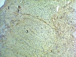
The collagen immunohystochemical typing in cervix of rats indicates the presence type III collagen therein. We have noted a weak positive expression of type III collagen in all of the layers in myometrium. (Fig. 1).
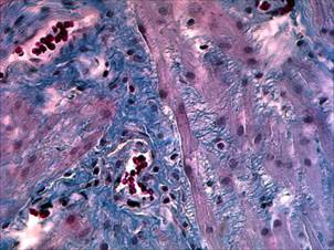
We found the increase of intercellular substance amount the labor before. The myocytes are isolated from each other. We found an interstitial edema seeing a light microscope (Fig. 2).
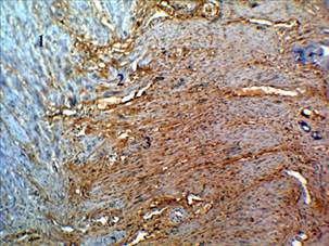
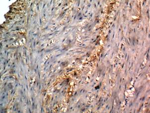
We discovered the increase of cervical collagen type III expression in labor (Fig. 3). This fact gives rise to an increase of its synthesis at the end of pregnancy and, especially, during labor. Noteworthy, different expression of collagen type III being studied in vascular and inner layers in comparison with outer layer. It also should be noted, that collagen type III is maximally visualized around myometrium vacuolated cells (Fig. 4). We found euchromatin dominance and presence of a 1-2 nucleoli is in nucleus in these cells viewing a light microscope. This fact suggests that secretion of type III collagen is carried by cervix leyomyocytes.
The literature report is controversial on this subject. There is evidence of reducing of collagen fibers density per unit area during pregnancy due to collagenolysis on the one hand [2,3,7]. There are some studies in which observed that the collagen does not change significantly on the other hand [5]. We think that myometrium collagen has a change from one type to another during labor. We see the formation of new young fibers of type III collagen.
Conclusion. We conclude in pregnancy and childbirth there are two interdependent processes: collagenolysis and collagenogenesis, growing at the time of delivery. The plastic function and integrity due cervix dilatation`s maintenance are realized by connective tissue during childbirth.The fundamental studies of structural changes at cellular and molecular levels in cervix require to develop the most effective methods of pregnancy and childbirth.
|
|
|
|
Fig. 1 Intact adult rat cervix /sphincter level. 1. The outer layer; 2. Vascular layer; 3. The inner circular layer. IHC typing collagen type III led. 40X.
|
Fig. 2 The rats cervix on day 19 of pregnancy. Vacuolated myocytes myometrium inner layer of the cervix. Colouring Masson. Zoom. 200X.
|
|
|
|
|
Fig. 3. The rat cervix during labor. 1. The outer layer; 2. Vascular layer; 3. The inner circular layer. IHC typing collagen type III led. 40X. |
Fig. 4. The rat cervix during labor. Positive expression of collagen around vacuolated myocytes. IHC typing of type III collagen. Zoom. 100 X. |
2. Buhimschi IA, Dussably L, Buhimschi CS, Ahmed A, Weiner CP. Physical and biomechanical characteristics of rat cervical ripening are not consistent with increased collagenase activity // Am J Obstet Gynecol. 2004 Nov;191(5):1695-704. PubMed PMID: 15547544.
3. Oxlund BS, Ørtoft G, Brüel A, Danielsen CC, Oxlund H, Uldbjerg N. Cervical collagen and biomechanical strength in non-pregnant women with a history of cervical insufficiency // Reprod Biol Endocrinol. 2010 Jul 30;8:92. doi: 10.1186/1477-7827-8-92.
4. Report of the AVMA Panel on Euthanasia // JAVMA.2001. Vol. 218, N. 5, Р. 669–696.
5. Resurrection SL The birth biomechanism: discrete-wave theory. Minsk: Polybius 1996 - 185 p.
6. Shkurupy VA, Myocytes different types dinamics in rat myometrium during pregnancy and the early postpartum involution. / VA Shkurupy, EV Dubinin, N. Dubinin // Bulletin of Experimental Biology and Medicine, 2008. - Annex 1 - From 101-104.
7. Uldbjerg N, Ekman G, Malmström A, Olsson K, Ulmsten U. Ripening of the human uterine cervix related to changes in collagen, glycosaminoglycans, and collagenolytic activity // Am J Obstet Gynecol. 1983 Nov 15;147(6):662-6.
Grigoryeva J. V., Suvorova G.N., Bormotov A.V., Chemidronov S.N. THE III TYPE COLLAGEN LOCALIZATION AND EXPRESSION FEATURES IN RATS UTERUS CERVIX DURING PREGNANCY AND CHILDBIRTH. International Journal Of Applied And Fundamental Research. – 2015. – № 1 –
URL: www.science-sd.com/460-24762 (24.04.2024).