About Us
Executive Editor:Publishing house "Academy of Natural History"
Editorial Board:
Asgarov S. (Azerbaijan), Alakbarov M. (Azerbaijan), Aliev Z. (Azerbaijan), Babayev N. (Uzbekistan), Chiladze G. (Georgia), Datskovsky I. (Israel), Garbuz I. (Moldova), Gleizer S. (Germany), Ershina A. (Kazakhstan), Kobzev D. (Switzerland), Kohl O. (Germany), Ktshanyan M. (Armenia), Lande D. (Ukraine), Ledvanov M. (Russia), Makats V. (Ukraine), Miletic L. (Serbia), Moskovkin V. (Ukraine), Murzagaliyeva A. (Kazakhstan), Novikov A. (Ukraine), Rahimov R. (Uzbekistan), Romanchuk A. (Ukraine), Shamshiev B. (Kyrgyzstan), Usheva M. (Bulgaria), Vasileva M. (Bulgar).
Medical sciences
Introduction
The problem of differentiation of various ophthalmic pathologies is still of importance. The major difficulties appear in diagnostics of inflammatory and dystrophic diseases of the posterior segment of the eyeball [1]. For example, choreoretinitis often have latent recurring character without any clinical symptoms. Standard diagnostic technique which is ophthalmoscopy is quite subjective. In addition the visual inspection of the fundus of eye is often complicated by a narrow rigid pupil, vitreous opacity and corneal dystrophy which make the diagnosis more uncertain. Similar alterations of retina and optical fiber may cover up various diseases: uncomplicated inflammatory processes, secondary retina dystrophy and exudative retinal detachment, optic neuritis, vitreous hemorrhage with formation of adhesions. Inflammatory focus in choroid at the first stage may be obscured by pigment epithelium detachment [2]. Study of functional disorders does not allow to increase diagnostic efficacy because both pathology types often cause identical alterations. Laboratory techniques also do not provide reliable differential diagnostics because alterations of biochemical and clinical parameters are low specific. Capabilities of additional instrumental diagnostics are also limited. Results of electrophysiological studies in this case are mostly non-specific and, consequently, not efficient. Rheoophatalmology which has been previously applied for such diagnostics primarily allows to evaluate hemodynamics of the anterior part of the uveal tract [3]. These circumstances significantly complicate clinical diagnostics.
In recent decade optical coherence tomography (OCT) has been successfully applied for diagnostics of eyeball pathologies [4]. OCT allows to obtain information about tissue pathologic alterations in all parts of eyeball [5, 6]. Possibility of repeating an OCT inspection allows to monitor the dynamics of the pathologic process and evaluate the efficacy of the chosen treatment procedure [7]. All this examples of OCT application in ophthalmology deal with non-contact diagnostics [4-7]. As for OCT diagnostics of anterior chamber angle (ACA) ciliary body (CB) pathologies, OCT is reputed to fail to provide information about ultrasructure of these objects and can be applied only for CB visualization in weak pigmented eyes [8].
OCT allows to reveal morphologic alterations while thermodiagnostics techniques have a potential to be used for evaluation of functional changes. Thermodiagnostics is a well-established technique which has found wide application in various medical areas [9-12]. Almost each disease is accompanied by changes in blood microcirculation and consequently, in thermoproduction. Thus, in the situation with inflammatory processes the increase in local temperature originates from increase in metabolism level, blood flow, and disorder in regulation of peripheral blood circulation. Accurate local temperature measurement may allow for differential diagnostics of various pathologies and provide monitoring of pathologic nidus evolution during treatment. Application of contact temperature measurement techniques has certain disadvantages such as the effect of contact quality on measurement accuracy.
The main goal of this paper is the demonstration of capability of contact OCT and non-contact IR-radiothermometry in diagnosing wide range of ophthalmic pathologies and illustration of the role of these technologies in ophthalmology.
Material and Methods
For contact OCT diagnostics we used OCT-1300U device (reg. certificate # 022а2005/235-05 May 5.2005) developed at the Institute of Applied Physics RAS (Nizhny Novgorod) with a specially developed en-face probe which is 2.7 mm in diameter. A supporting beam in the visible range (630 nm, 0.1 mW), which indicates the scanning direction at the sample surface is integrated into the scanning system. Principal scheme and technical characteristics of the setup are described in other papers [13].
The developed microprocessor IR radiothermometer [14,15] is based on the scheme of modulation radiometer (Fig.1).

Fig. 1. Structural diagram of microprocessor radiometer
The radiometer utilizes the modulation measurement technique. It is based on the periodic connection of the measured signal proportional to the temperature of the studied object and reference noise source with known temperature proportional to the input channel of the radiometer. Trigging is performed by employment of reference voltage generator with meander-shaped voltage time dependence. The essential feature of the developed device is the modulation of radiation in front of two-channel optical system with a diaphragm [13], which allows to avoid errors connected with possibility of measurement of the optical system temperature. Absolute error of measurement does not exceed 0.05°С, size of focal spot is 5 mm, the wavelength range is from 2 to 25 mkm.
At the stage of primary diagnostics all the patients involved in the study have undergone full set of standard inspection procedures.
In the framework of this study, 34 healthy volunteers (68 eyes), 72 patients with eye contusion of different grade (144 eyes), 5 eyes ex vivo and 15 eyes post mortem were inspected with OCT for possible comparison of OCT and histologic data. OCT diagnostics in vivo was performed under local instillation anesthesia with 1% dicain solution. The OCT probe contact surface was fixed on bulbar conjunctiva.
In the IR radiometry study we have inspected 49 healthy volunteers, 54 patients with various dystrophic diseases of the posterior segment of the eye, and 47 patients with inflammatory processes at fundus (choreoretinitis, optic neuritis). The inspection was performed without anesthesia because the technique is non-contact. For minimization of measurement errors related to the influence of environmental conditions reference measurements are used in IR thermometry. Registration of temperature was performed both in the pathology area and in the symmetric one, i.e. the temperature was measured consecutively in both eyes of the patient and then the measured values were compared. The measurements were performed two or three times in order to minimize the measurement error. The examination of one patient with this method took only 1.0 - 1.5 minutes, which could be useful in the outpatient setting.
Results
Typical OCT images of the anterior segment of the eyeball in visualization aimed for corner of anterior chamber and ciliary body were obtained from healthy patients. In 75 % of OCT-images obtained in 1 mm from limb it is possible to distinguish histologic structures of the drainage system: anterior chamber corner, trabecular apparatus and Schlemm's canal (Fig.2a, b). Trabecular apparatus appeared in OCT-images as a non-uniform structure with prevailing high signal level and pronounced continuous irregular boundaries. Schlemm's canal appeared as low scattering convex area with continuous irregular contrasted boundaries..
a 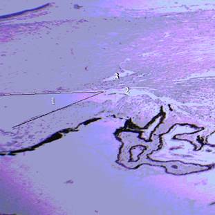 ь
ь 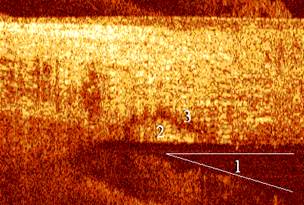
c 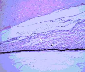 d
d 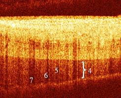
Fig. 2. Histology of anterior chamber corner and ciliary body section in 1 mm (a) and 2 mm (c) from limb; OCT-images of normal anterior chamber corner section in 1 mm (b) and 2 mm (d) from limb
1-anterior chamber corner; 2- trabecular apparatus; 3- Schlemm's canal; 4- ciliated part of CB; 5-muscular layer of CB; 6-vascular layer of CB; 7- pigment epithelium)
In 90% of OCT-images in 2 mm from limb the ciliated part of CB appeared as a triangle with truncated apex corresponding to histological transversal sections of the ciliated part of CB (Fig.2c,d). In the OCT-images of the ciliated part of CB we differentiated the main layers. The muscular layer appeared as a structure with moderate OCT signal level and regular contrasted boundaries. The vascular layer of CB appeared as an area with lowered signal and irregular weak boundaries. The pigment epithelium was manifested in the form of high signal level and irregular contrasted boundaries and regular or scalloped lower boundary. We did not find any dependence of the OCT appearance of these structures on age or gender which allowed to use these images as reference for non-altered structures of the anterior chamber.
In order to develop criteria for recognition of altered structures of the anterior segment in OCT-images we performed OCT-diagnostics of patients with different grade of eye contusion. This study discovered a set of typical morphologic changes in the anterior segment of eye manifested in OCT-images. We have discovered that the earliest and essential pathomorphological manifestation of the eyeball contusion is the edema, while its stage and localization depend on the grade. We have found several variants of manifestation of edema of the anterior segment inner structures: thickening of one or more layers of CB, the appearance of zones with reduced signal level of irregular shape without distinguished boundaries in the muscular and vascular layers, disturbance of CB layers visualization in an OCT-image with extreme manifestation of structure loss in the image, which is typical for severe concussion of the eyeball (Fig. 3).
a 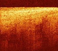 b
b 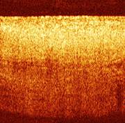
Fig. 3. Typical OCT images of CB with severe concussion of the eyeball: typical structure loss with presence of chaotic areas with low signal level
The IR radiothermometry has been employed for differential diagnostics of dystrophic and inflammatory diseases of the eyeball. The results show that dystrophic processes of the posterior segment of the eyeball are accompanied by decrease of the surface temperature by a value from 0.3 to 1.2 °С. Inflammatory diseases of the posterior segment of the eyeball are accompanied by increase of the surface temperature by a value from 0.3 to 0.8 °С. It is worth mentioning that the temperature variation value depends on the grade of a pathologic process.
 >
>
Fig. 4. Temperature asymmetry of the anterior and posterior segments of the eye obtained from IR radiothermometry measurements (grey bars show confidence intervals of the measurements, dash lines show the interval for average norm values). 1 - norm; 2 – dystrophic diseases of the anterior segment of the eye (corneal dystrophy); 3 - inflammatory diseases diseases of the anterior segment of the eye (keratitis and iridocyclitis); 4 - dystrophic diseases of the posterior segment of the eye (of retina and optic nerve); 5 – inflammatory diseases of the posterior segment of the eye
Based on the results of the reference group we found that without eye pathology the temperature asymmetry between two eyes does not exceed 0.1 – 0.25 °С. Pathology of the eyeball is accompanied by the revealed pronounced temperature asymmetry which allows to perform differential diagnostics of inflammatory and dystrophic processes of the eyeball.
Fig. 4 shows histograms of temperature asymmetry for pathologies of anterior and posterior eye segments. The results show that the temperature asymmetry has different character for inflammatory and dystrophic diseases, in pathologies of the posterior segment it is much lower compared to diseases of the anterior segment. This result is natural and is explained by deeper location of the thermal inhomogeneity in pathologies of the posterior segment.
Against the background of anti-inflammatory and immunocorrective therapy in inflammatory patients the temperature asymmetry decreased (Fig. 5) and finally became undetectable which can be considered as an additional criterion of remission
 >
>
Fig. 5. Evolution of temperature asymmetry during treatment of acute uveitis (1), torpid uveitis (2) and senile macular degeneration (3)
Discussion and conclusion
In this paper we discuss two diagnostic techniques which are being actively developed in the recent decade in various medical areas including ophthalmology. OCT has already become a standard procedure in eyeball pathologies diagnostics. More than that, Anterior Segment Optical Coherence Tomography (AS-OCT) technique has been developed. The AS-OCT is a non-contact, non-invasive imaging modality that provides high resolution image of the anterior segment in cross section in vivo [16,17]. Furthermore the use of wide-field scanning optics (16 mm) and deep axial scan range (8 mm) allows the AS-OCT to image the entire anterior chamber in a single frame. It allows for an objective assessment of the anterior chamber (AC) angle and is easy to use even with minimal training. The considered modality of contact ОСТ provides possibility not only for diagnostics of the anterior chamber corner which is important for glaucoma diagnostics, but also for imaging of inner structures of the anterior chamber, ciliary body and Schlemm's canal. This provides novel capabilities for OCT application in ophthalmology. Combination of anterior chamber corner evaluation and imaging of Schlemm's canal shape is promising not only for glaucoma diagnostics, but also for differential diagnostics of various forms of this pathology. In this paper we show that contact OCT can be employed for revealing of the grade of the eyeball contusion. Comparison of the results of OCT diagnostics for patients of three groups with contusions of different grade demonstrated statistically significant difference between the groups by OCT-criteria. It is important that statistically significant difference can be observed in OCT-criteria at different stages of (early and delayed manifestations, convalescence period). This allows to employ OCT not only for diagnostics, but also for monitoring of treatment of eye contusion.
In this paper we show that non-contact IR radiothermometry is a convenient tool both for conventional and differential diagnostics, as well as for monitoring of treatment of different eyeball pathologies.
In our opinion there is no need to compare the applied techniques because they are different both in principle of operation and information obtained. However, combination of these techniques will allow to increase diagnostic efficacy: OCT allows to monitor evolution of morphologic (patomorphologic) alterations at the level close to histology while IRRT allows to monitor tissue temperature. Radiothermometry can be combined both with conventional OCT and contact OCT considered in this work. For example, combination of non-contact OCT and IRRT is very promising for monitoring of structure and temperature of the posterior eye segment in transpupillary thermotherapy, which is applied both in tumor [18], and non-tumor eye pathologies [19].
In conclusion we should point out that the considered in the paper OCT and IRRT techniques are non-invasive and safe diagnostic techniques that provide possibility for multiple application monitoring and follow-up. For IRRT non-invasiveness is an additional advantage, however, contactness of OCT in the considered modification provides advantages in visualization of anterior segment structures. Both techniques are promising for diagnostics of wide range of eye pathologies (inflammatory processes, dystrophic diseases, contusion, glaucoma). They could be employed both for primary and differential diagnostics at the early or delayed stage. Their combination is promising for evaluation of morphologic and functional state of the eyeball.
Acknowledgements
This work was supported by the Government of the Russian Federation, Grant No. 11.G34.31.0066.
References
- Telander DG. Inflammation and age-related macular degeneration (AMD). Semin Ophthalmol. 2011 May;26(3):192-7
- Donoso LA, Kim D, Frost A, Callahan A, Hageman G. The role of inflammation in the pathogenesis of age-related macular degeneration. Surv Ophthalmol. 2006 Mar-Apr;51(2):137-52
- Bressler N.M. For the treatment of macular degeneration – Tap report 1. Arch. Ophtalmol. 117/1329-1345, 1999
- Optical Coherence Tomography of Ocular Diseases, Schuman JS, Puliafito CA, Fujimoto JG, Duker JS, eds.Third Edition, 2013, SLACK Incorporated 640 pp
- Andre J. Witkin, MD; Nikolas J. S. London, MD; Jonathan D. Wender, MD; Arthur Fu, MD; Sunir J. Garg, MD; Carl D. Regillo, MD// Spectral-Domain Optical Coherence Tomography of White Dot Fovea. Arch Ophthalmol. 2012;130(12):1603-1605
- Reis AS , Sharpe GP , Yang H , et al. Optic disc margin anatomy in patients with glaucoma and normal controls with spectral domain optical coherence tomography. Ophthalmology . 2012;119:738–747
- Massin P, Allouch C, Haouchine B, Metge F, Paques M, Tangui L, Erginay A, Gaudric A. Optical coherence tomography of idiopathic macular epiretinal membranes before and after surgery. Am J Ophthalmol 2000; 130: 732-739
- Baikoff G, Lutun E, Ferraz C, Wei J (2004) Static and dynamic of the Anterior Segment with Optical Coherence Tomography. J Cataract Refract Surg (Sept 2004) Vol. 30 No.9 pp. 1843-50
- Taberner A.J., Hunter I.W., Kirton R.S., Nielsen P.M.F., Loiselle D.S. Characterization of a flowthrough microcalorimeter for measuring the heat production of cardiac trabeculae // Review of Scientific Instruments. - 2005. - V.76, Is.10. - P.104902
- Hillery M.T., Shuaib I. Temperature effects in the drilling of human and bovine bone // Journal of Materials Processing Technology. - 1999. - V.92-93. - P.302-308
- Rosenthal H.M., Leslie A. Measuring temperature of NICU patients – A comparison of three devices. J. Neonatal Nurs. – 2006, 12, 125-129
- Verkruyssc W, Jia W, Franco W, Milner ТЕ, Nelson JS. Infrared measurement of human skin temperature to predict the individual maximum safe radiant exposure (IMSRE).// Lasers Surg Med. 2007 Dec., 39(10):757-66
- Dolin L.S., Feldchtein F.I., Gelikonov G.V., Gelikonov V.M., Gladkova N.D., Iksanov R.R., Kamensky V.A., Kuranov R.V., Sergeev A.M., Shakhova N.M., Turchin I.V. Fundamentals and Clinical Applications of the PM-Fiber Based Endoscopic OCT // Coherent-Domain Optical Methods Biomedical Diagnostics, Environmental and Material Science./: Kluwer Academic Publishers, 2004. - P.- 211-271.
- Orlov I.Ya. Switching Radiometer of Infrared Radiation / Afanas’ev A.V., Nikiforov I.A., Orlov P.I., Terent’ev I.G. // G01 J5/10 (2006.01) RF Patent № 2345333, 23.08.07
- Orlov I.Ya., Afanas’ev A.V., Nikiforov, I.A. High-precision Radiometer of Infrared Radiation// Automation Remote Control, 2011, v. 72, № 4, p.345-349
- Leung CK, Cheung CY, Li H, Dorairaj S, Yiu CK, Wong AL, Liebmann J, Ritch R, Weinreb R & Lam DS. Dynamic Analysis of Dark–Light Changes of the Anterior. Chamber Angle with Anterior Segment OCT Invest Ophthalmol Vis Sci., Vol. 48, No, 2007, pp. 4116–4122
- Liu L. Anatomical Changes of the Anterior Chamber Angle With Anterior-Segment Optical Coherence Tomography Arch Ophthalmol., 2008, Vol. 126, No. 12, pp.1682-1686
- Shields CL, Shields JA, Perez N, et al.: Primary transpupillary thermotherapy for small choroidal melanoma in 256 consecutive cases: outcomes and limitations. Ophthalmology 109 (2): 225-34, 2002
- Newsom, R. S., McAlister, J. C., Saeed, M., and McHugh, J. D. Trathermotherapy (TTT) for the treatment of choroidal neovascularisation. British Journal of Ophthalmology 2001; 85: 173-178
Pavel Orlov, Georgy Bogdanov, Igor Orlov, Valentin Gelikonov and Natalia Shakhova CONTACT OPTICAL COHERENCE TOMOGRAPHY AND NON-CONTACT IR RADIOTHERMOMETRY IN DIAGNOSING NON-TUMOR PATHOLOGIES IN OPHTALMOLOGY . International Journal Of Applied And Fundamental Research. – 2013. – № 1 –
URL: www.science-sd.com/452-24044 (11.02.2026).


 PDF
PDF